Understanding Endoscopic Spine Imaging: A Technological Marvel in Modern Manufacturing
Endoscopic Spine Imaging is a state-of-the-art, minimally invasive, diagnostic and therapeutic procedure that allows surgeons to diagnose and treat patients with spinal disorders, without the need for large incisions. This innovative technology has revolutionized the world of spinal surgery, making it easier for physicians and patients alike to achieve optimal health outcomes. Let's explore how Endoscopic Spine Imaging works and understand its key components and working mechanisms.
How Endoscopic Spine Imaging Works:
The procedure involves the use of an endoscope, which is a long, slender tube equipped with a camera, light, and lens. This endoscope is inserted through small incisions, into the spine, and relays video images of the spine to a monitor. Endoscopic Spine Imaging requires anesthesia and is usually performed as an outpatient procedure, allowing patients to go home the same day.
Key Components and Working Mechanisms:
One of the key components of Endoscopic Spine Imaging is the endoscope, a slender tube that is inserted through small incisions into the spinal canal. The endoscope carries a camera, light, and lens, which allows the surgeon to see inside the spine, where traditional imaging techniques cannot reach. A monitor displays the images captured by the endoscope.
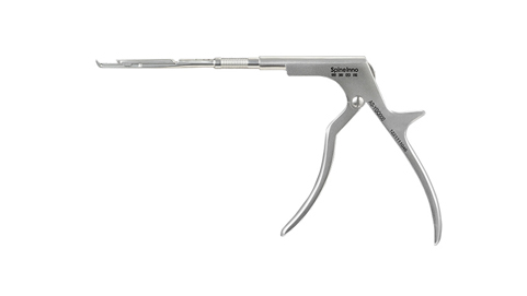
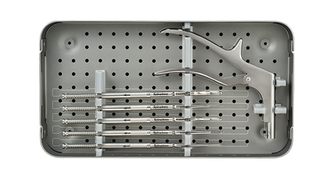
Another key component is the specialized surgical tools, which are used to perform various procedures through the endoscope. These tools are designed to deliver precision and accuracy, while minimizing damage to surrounding tissue and nerves. Some procedures that can be performed through Endoscopic Spine Imaging include discectomy, laminectomy, and fusion.
Advanced Technology and Efficient Production Processes:
Endoscopic Spine Imaging is a product of advanced technology and has been instrumental in providing modern manufacturing efficiency that has become increasingly important in modern manufacturing. The technology has enabled efficient production processes in the manufacturing of the endoscopes and surgical tools, guaranteeing their quality and minimizing the risk of malfunctions during surgical procedures.
Application Scenarios:
In practice, Endoscopic Spine Imaging has helped thousands of patients with spine disorders. For instance, for patients with herniated disks, Endoscopic Spine Imaging is used to view and remove the herniated portions without damaging surrounding spinal tissue. For patients with spinal stenosis, Endoscopic Spine Imaging is used to visualize and remove bone spurs that are causing nerve compression.
Conclusion:
In conclusion, Endoscopic Spine Imaging is a cutting-edge technology that offers minimally invasive diagnostic and therapeutic procedures for patients with spinal disorders. The key components of this technology include the endoscope, specialized surgical tools, and advanced video imaging, which enable surgeons to achieve precision and accuracy in their procedures. Endoscopic Spine Imaging has revolutionized modern manufacturing by enabling efficient production processes and has become an important part of modern healthcare. For inquiries on Endoscopic Spine Imaging, Contact us, and our suppliers will be glad to assist you.


 Spine Specialist